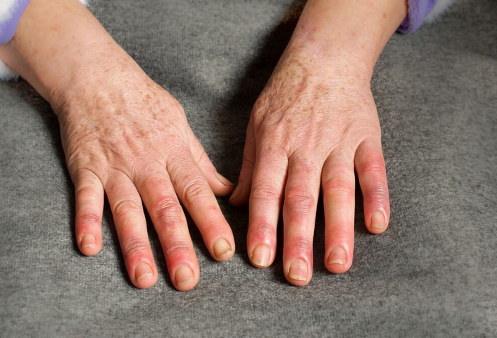
Torn Chest Muscle: Causes, Symptoms, and Recovery

Source : https://paininjuryrelief.com

A torn chest muscle, medically known as a pectoral muscle strain or rupture, is a condition that occurs when the tendons or muscles of the pectoral (chest) region are stretched or torn. This can be caused by various factors such as excessive weightlifting, sudden movements, or direct blows to the area. Symptoms often include pain, swelling, bruising, and a decrease in chest muscle strength. Recovery from a torn chest muscle involves rest, physical therapy, and in severe cases, surgical intervention. It is a process that requires time and patience, with the focus on gradually restoring strength and function to the affected area.

Understanding Torn Chest Muscle: Causes, Symptoms, and Steps to Recovery

A torn chest muscle, medically known as a pectoralis major rupture, is a severe injury that can significantly impact an individual’s quality of life. Understanding the causes, symptoms, and steps to recovery can help those affected by this condition navigate their healing journey more effectively.

The pectoralis major is a large muscle in the upper chest, responsible for controlling arm movements such as pushing and lifting. A tear in this muscle typically results from excessive strain or a sudden, forceful movement. This injury is most common among athletes, particularly those involved in weightlifting, wrestling, and football, where intense physical exertion and contact are frequent. However, it can also occur in non-athletes due to accidents or falls.

The symptoms of a torn chest muscle are usually immediate and noticeable. The individual may experience a sudden sharp pain in the chest or upper arm, often described as a tearing or popping sensation. This may be accompanied by swelling, bruising, and a noticeable deformity in the chest muscle. The pain can intensify with arm movement, making it difficult to perform daily activities. In some cases, the individual may also experience muscle weakness or an inability to move the arm across the body.

Diagnosis of a torn chest muscle typically involves a physical examination and imaging tests such as an MRI or ultrasound. These tests can help determine the severity of the tear and guide the treatment plan. It’s crucial to seek medical attention promptly if a pectoralis major rupture is suspected, as delayed treatment can lead to complications and prolonged recovery.

The recovery process for a torn chest muscle depends on the severity of the injury. Mild to moderate tears may heal with conservative treatment, including rest, ice, compression, and elevation (RICE). Pain medication and physical therapy may also be recommended to manage pain and restore strength and flexibility to the muscle.

In the case of severe tears, surgical intervention may be necessary. This typically involves reattaching the torn muscle to the bone using sutures. Post-surgery, the individual will likely need to wear a sling for several weeks to immobilize the arm and allow the muscle to heal. Physical therapy will then be introduced gradually to regain muscle strength and range of motion.

Regardless of the treatment approach, recovery from a torn chest muscle requires time and patience. The healing process can take several months, and it’s essential to avoid rushing back into physical activities to prevent re-injury. Regular follow-ups with the healthcare provider are also crucial to monitor progress and adjust the treatment plan as needed.

In conclusion, a torn chest muscle is a serious injury that requires prompt medical attention. Understanding the causes and symptoms can help individuals seek timely treatment, while knowledge of the recovery process can set realistic expectations and promote effective healing. With the right care and commitment to recovery, individuals with a torn chest muscle can regain their strength and return to their regular activities.A torn chest muscle, also known as a pectoral muscle rupture, is often caused by excessive strain or injury during activities such as weightlifting or contact sports. Symptoms typically include sudden severe pain, swelling, bruising, muscle weakness, and a noticeable deformity in the chest muscle. Recovery involves rest, pain management, physical therapy, and in severe cases, surgical intervention. The healing process can be lengthy, often requiring several weeks to months. It is crucial to seek immediate medical attention if a chest muscle tear is suspected to ensure proper treatment and recovery.

Swollen Finger Joint: Common Causes and Treatment

Source : https://www.geelongmedicalgroup.com.au

Swollen Finger Joint refers to the inflammation or enlargement of the joints in the fingers, often resulting in pain, stiffness, or discomfort. This condition can be caused by various factors including injuries, infections, arthritis, gout, or other underlying health conditions. Treatment typically involves managing the symptoms through medication, physical therapy, lifestyle changes, or in severe cases, surgery. It’s crucial to consult a healthcare professional for an accurate diagnosis and appropriate treatment plan.

Understanding Swollen Finger Joint: Common Causes and Treatment

Swollen finger joints can be a common occurrence, often causing discomfort and hindering daily activities. Understanding the causes and treatment options for this condition can help individuals manage the symptoms and seek appropriate medical attention when necessary.

One of the most common causes of a swollen finger joint is arthritis, a condition characterized by inflammation of the joints. There are several types of arthritis, including osteoarthritis and rheumatoid arthritis, both of which can lead to swelling in the finger joints. Osteoarthritis typically occurs due to wear and tear on the joints over time, while rheumatoid arthritis is an autoimmune disorder where the body’s immune system mistakenly attacks its own tissues, including the joints.

Another common cause of swollen finger joints is an injury or trauma to the hand. This could be a result of a fall, a sports injury, or even repetitive strain from activities such as typing or playing a musical instrument. In these cases, the swelling is often accompanied by pain and may be temporary, subsiding as the injury heals.

Infections can also lead to swelling in the finger joints. For instance, a condition known as septic arthritis occurs when a bacterial or fungal infection enters a joint, leading to inflammation and swelling. This condition requires immediate medical attention as it can quickly lead to joint damage if left untreated.

Gout, a form of arthritis caused by excess uric acid in the body, can also cause swollen finger joints. In this condition, the excess uric acid forms crystals in the joints, leading to intense pain and swelling. Gout attacks can occur suddenly and often at night.

Treatment for a swollen finger joint largely depends on the underlying cause. For arthritis-related swelling, treatment may involve medication to reduce inflammation and pain, as well as physical therapy to improve joint function. Lifestyle changes, such as maintaining a healthy weight and regular exercise, can also help manage symptoms.

In the case of an injury, treatment may involve rest, ice, compression, and elevation (RICE) to reduce swelling and promote healing. Over-the-counter pain relievers may also be recommended. If the injury is severe, medical intervention such as surgery may be necessary.

For infections like septic arthritis, antibiotics are typically prescribed to fight the infection. In some cases, surgical drainage of the infected joint may be required.

Gout is usually treated with medications to control uric acid levels and reduce inflammation. Dietary changes, such as reducing intake of foods high in purines, can also help manage gout.

In conclusion, a swollen finger joint can be caused by a variety of conditions, from arthritis to injuries to infections. Treatment options vary depending on the cause, but often involve medication, lifestyle changes, and in some cases, surgery. If you’re experiencing a swollen finger joint, it’s important to seek medical attention to determine the cause and appropriate treatment. Remember, early intervention can help prevent further joint damage and improve your quality of life.Swollen finger joints can be caused by a variety of conditions, including arthritis, gout, injury, infection, or certain diseases like lupus. The treatment typically involves addressing the underlying cause, which may include medication, physical therapy, lifestyle changes, or in severe cases, surgery. It’s important to consult with a healthcare professional for an accurate diagnosis and appropriate treatment plan.

Painful Pectoral Muscle: Causes, Symptoms, and Recovery Strategies

Painful pectoral muscle is a condition that affects the chest muscles, causing pain and discomfort. It can be caused by a variety of factors, including overuse, injury, or medical conditions. Symptoms of painful pectoral muscle can include pain in the chest, difficulty breathing, and difficulty moving the arms. Treatment for painful pectoral muscle typically involves rest, physical therapy, and medications. Recovery strategies may include stretching, strengthening exercises, and lifestyle modifications. This article will discuss the causes, symptoms, and recovery strategies for painful pectoral muscle.

Understanding the Causes of Painful Pectoral Muscles: What You Need to Know

Painful pectoral muscles can be a source of discomfort and frustration for many people. Understanding the causes of this condition can help you take steps to reduce your pain and improve your quality of life. In this article, we’ll discuss the potential causes of painful pectoral muscles and what you can do to manage your symptoms.

The most common cause of painful pectoral muscles is overuse. This can occur when you engage in activities that require repetitive motions, such as weightlifting or swimming. Overuse can also occur when you perform activities that require a lot of force, such as pushing or pulling heavy objects. In addition, poor posture can lead to overuse of the pectoral muscles, as can poor technique when performing exercises.

Injury is another potential cause of painful pectoral muscles. This can occur when you experience a sudden, forceful movement that causes a strain or tear in the muscle. This type of injury can be caused by a fall, a car accident, or a sports-related incident.

In some cases, painful pectoral muscles can be caused by an underlying medical condition. This can include conditions such as arthritis, bursitis, or tendonitis. It can also be caused by an infection or an autoimmune disorder.

Finally, it’s important to note that some medications can cause painful pectoral muscles. These include certain types of antibiotics, steroids, and chemotherapy drugs.

If you’re experiencing painful pectoral muscles, it’s important to talk to your doctor. They can help you determine the cause of your pain and recommend the best treatment plan for you. Treatment may include rest, physical therapy, medications, or surgery.

In addition to seeking medical treatment, there are some things you can do at home to help reduce your pain. These include avoiding activities that cause pain, stretching regularly, and using heat or cold therapy. You should also make sure to practice good posture and use proper technique when performing exercises.

By understanding the causes of painful pectoral muscles, you can take steps to reduce your pain and improve your quality of life. If you’re experiencing pain, talk to your doctor to determine the best treatment plan for you.

Exploring the Symptoms of Painful Pectoral Muscles: How to Identify and Treat

Painful pectoral muscles can be a source of discomfort and can interfere with daily activities. It is important to identify the cause of the pain and to seek appropriate treatment. This article will discuss the symptoms of painful pectoral muscles, how to identify them, and how to treat them.

Symptoms of Painful Pectoral Muscles

Painful pectoral muscles can cause a variety of symptoms, including:

• Pain in the chest area, which may be localized or spread out
• Tenderness or soreness in the chest area
• Difficulty moving the arms or shoulders
• Weakness in the arms or shoulders
• Swelling or inflammation in the chest area
• Difficulty breathing

How to Identify Painful Pectoral Muscles

If you are experiencing any of the above symptoms, it is important to seek medical attention. Your doctor will be able to diagnose the cause of your pain and recommend the best course of treatment.

Your doctor may order imaging tests, such as an X-ray or MRI, to help diagnose the cause of your pain. They may also order blood tests to check for any underlying medical conditions that could be causing the pain.

How to Treat Painful Pectoral Muscles

Once the cause of your pain has been identified, your doctor will recommend the best course of treatment. Treatment options may include:

• Rest: Resting the affected area can help reduce pain and inflammation.

• Ice: Applying ice to the affected area can help reduce swelling and pain.

• Medication: Over-the-counter or prescription medications may be recommended to help reduce pain and inflammation.

• Physical therapy: A physical therapist can help you strengthen the affected muscles and improve range of motion.

• Surgery: In some cases, surgery may be necessary to repair any damage to the muscles or tendons.

Conclusion

Painful pectoral muscles can be a source of discomfort and can interfere with daily activities. It is important to identify the cause of the pain and to seek appropriate treatment. This article has discussed the symptoms of painful pectoral muscles, how to identify them, and how to treat them. If you are experiencing any of the above symptoms, it is important to seek medical attention. With the right diagnosis and treatment, you can reduce your pain and improve your quality of life.In conclusion, painful pectoral muscle can be caused by a variety of factors, including overuse, injury, and medical conditions. Symptoms of this condition can range from mild to severe and may include pain, tenderness, and difficulty moving the affected area. Treatment options vary depending on the cause of the pain, but may include rest, physical therapy, medications, and surgery. Recovery strategies such as stretching, strengthening exercises, and proper nutrition can help to reduce pain and improve overall function.

Swollen Knuckle: Causes, Treatment, and When to See a Doctor

Swollen knuckles can be a painful and uncomfortable condition that can affect anyone at any age. It is caused by a variety of factors, including injury, arthritis, and infection. Treatment for swollen knuckles depends on the underlying cause, but may include rest, ice, compression, and elevation. In some cases, medications or surgery may be necessary. It is important to seek medical attention if the swelling does not improve or if it is accompanied by other symptoms. This article will discuss the causes, treatment, and when to see a doctor for swollen knuckles.

What Causes Swollen Knuckles and How Can You Treat Them?

Swollen knuckles, also known as Heberden’s nodes, are a common symptom of osteoarthritis, a degenerative joint disease. Osteoarthritis is caused by the breakdown of cartilage, the tissue that cushions the ends of bones in a joint. As the cartilage wears away, the bones rub against each other, causing pain, swelling, and stiffness.

There are several treatments available for swollen knuckles. The most important step is to reduce the inflammation and pain. This can be done with over-the-counter anti-inflammatory medications such as ibuprofen or naproxen. Applying ice to the affected area can also help reduce swelling and pain.

Physical therapy can also be beneficial in treating swollen knuckles. Exercises that focus on strengthening the muscles around the joint can help reduce pain and improve range of motion. Stretching exercises can also help reduce stiffness.

In some cases, a doctor may recommend a corticosteroid injection to reduce inflammation and pain. Surgery may also be an option for severe cases of osteoarthritis.

It is important to talk to your doctor about the best treatment option for your swollen knuckles. With the right treatment, you can reduce pain and improve your quality of life.

When Should You See a Doctor for Swollen Knuckles?

If you experience swollen knuckles, it is important to seek medical attention as soon as possible. Swollen knuckles can be a sign of a serious medical condition, such as arthritis, gout, or an infection. It is important to have your knuckles examined by a doctor to determine the cause of the swelling and to receive the appropriate treatment.

If you experience any of the following symptoms in addition to swollen knuckles, it is especially important to seek medical attention right away: redness, warmth, pain, tenderness, or difficulty moving the joint.

If you are experiencing swollen knuckles, it is important to make an appointment with your doctor as soon as possible. Your doctor will be able to diagnose the cause of the swelling and provide you with the appropriate treatment.Swollen knuckles can be caused by a variety of conditions, including arthritis, gout, and trauma. Treatment for swollen knuckles depends on the underlying cause and may include rest, ice, compression, elevation, and medications. If the swelling persists or is accompanied by other symptoms, it is important to see a doctor for a proper diagnosis and treatment plan.

Torn Pectoral Muscle: Causes, Symptoms, and Recovery Strategies

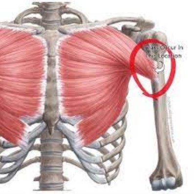
A torn pectoral muscle is a serious injury that can cause significant pain and disability. It is most commonly seen in athletes who participate in activities that involve pushing or pulling motions, such as weightlifting, football, and wrestling. The pectoral muscle is a large muscle located in the chest that helps to move the arm and shoulder. When it is torn, it can cause pain, swelling, and difficulty moving the arm. Treatment for a torn pectoral muscle typically involves rest, physical therapy, and possibly surgery. This article will discuss the causes, symptoms, and recovery strategies for a torn pectoral muscle.

Understanding the Causes of a Torn Pectoral Muscle: What You Need to Know

A torn pectoral muscle is a serious injury that can cause significant pain and limit mobility. Understanding the causes of a torn pectoral muscle can help you prevent this injury and seek appropriate treatment if it does occur.

The pectoral muscles are located in the chest and are responsible for moving the arms and shoulders. A tear in the pectoral muscle can occur due to a sudden, forceful movement of the arm or shoulder, such as when lifting a heavy object or performing a strenuous activity. It can also occur due to repetitive strain, such as when performing the same motion over and over again.

In addition to sudden or repetitive strain, a torn pectoral muscle can also be caused by a direct blow to the chest. This can occur during contact sports, such as football or rugby, or during a car accident.

The symptoms of a torn pectoral muscle include pain in the chest, shoulder, and arm, as well as swelling and bruising. You may also experience difficulty moving your arm or shoulder, and you may hear a popping sound when the injury occurs.

If you suspect that you have a torn pectoral muscle, it is important to seek medical attention as soon as possible. Your doctor will be able to diagnose the injury and recommend a course of treatment. Treatment may include rest, ice, compression, and elevation, as well as physical therapy and medications to reduce pain and inflammation. In some cases, surgery may be necessary to repair the torn muscle.

By understanding the causes of a torn pectoral muscle, you can take steps to prevent this injury. Make sure to warm up before engaging in any strenuous activity, and avoid lifting heavy objects without proper form. If you do experience any pain or discomfort in your chest, shoulder, or arm, seek medical attention right away.

Diagnosing and Treating a Torn Pectoral Muscle: Symptoms and Recovery Strategies

A torn pectoral muscle can be a painful and debilitating injury. It is important to recognize the symptoms of a torn pectoral muscle and to seek medical attention as soon as possible. With proper diagnosis and treatment, a full recovery is possible.

Symptoms

The most common symptom of a torn pectoral muscle is sudden, sharp pain in the chest area. This pain may be accompanied by swelling, bruising, and difficulty moving the arm. Other symptoms may include a popping sensation in the chest, numbness, and weakness in the arm.

Diagnosis

If you suspect you have a torn pectoral muscle, it is important to seek medical attention. Your doctor will likely perform a physical exam and ask questions about your symptoms. Imaging tests such as an X-ray or MRI may also be used to confirm the diagnosis.

Treatment

Treatment for a torn pectoral muscle will depend on the severity of the injury. In mild cases, rest and physical therapy may be enough to promote healing. In more severe cases, surgery may be necessary to repair the muscle.

Recovery Strategies

Once the injury has been treated, it is important to follow a rehabilitation program to ensure a full recovery. This may include physical therapy, stretching, and strengthening exercises. It is also important to rest and avoid activities that could aggravate the injury.

Conclusion

A torn pectoral muscle can be a painful and debilitating injury. It is important to recognize the symptoms and seek medical attention as soon as possible. With proper diagnosis and treatment, a full recovery is possible with the help of a rehabilitation program.Torn pectoral muscles can be a painful and debilitating injury, but with proper diagnosis and treatment, recovery is possible. Physical therapy, rest, and proper nutrition are all important components of a successful recovery. It is important to follow the advice of your doctor and physical therapist to ensure a safe and successful recovery. With the right care and attention, you can return to your normal activities and enjoy a full and healthy life.

Torn Pec Recovery: Rehabilitation and Healing After a Pectoral Muscle Injury

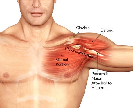
Torn Pec Recovery is a comprehensive guide to rehabilitation and healing after a pectoral muscle injury. It provides detailed information on the causes, diagnosis, and treatment of pectoral muscle injuries, as well as advice on how to prevent them in the future. It also offers practical tips on how to manage the pain and discomfort associated with a pectoral muscle injury, as well as how to speed up the healing process. With the help of Torn Pec Recovery, you can get back to your active lifestyle in no time.

Understanding the Anatomy of the Pectoral Muscle and How to Best Recover from a Torn Pec Injury

The pectoral muscles, commonly referred to as the “pecs,” are a group of muscles located in the chest area. They are responsible for a variety of movements, including flexing the shoulder, rotating the arm, and adducting the arm. The pectoral muscles are also important for providing stability to the shoulder joint.

Unfortunately, the pectoral muscles are prone to injury, particularly tears. A torn pectoral muscle can be a very painful and debilitating injury. It is important to understand the anatomy of the pectoral muscles and the best ways to recover from a torn pectoral muscle injury.

The pectoral muscles are made up of two main muscles: the pectoralis major and the pectoralis minor. The pectoralis major is the larger of the two muscles and is responsible for most of the movement of the shoulder joint. It originates from the sternum and clavicle and inserts into the humerus. The pectoralis minor is a smaller muscle located underneath the pectoralis major. It originates from the ribs and inserts into the shoulder joint.

Tears of the pectoral muscles can occur due to a variety of causes, including overuse, trauma, or sudden force. Symptoms of a torn pectoral muscle include pain, swelling, and bruising in the chest area. It is important to seek medical attention if you suspect you have a torn pectoral muscle.

The best way to recover from a torn pectoral muscle injury is to rest and allow the muscle to heal. It is important to avoid any activities that may cause further injury or strain the muscle. Ice and anti-inflammatory medications can help reduce pain and swelling. Physical therapy can also be beneficial in helping to restore strength and range of motion.

Surgery may be necessary in some cases of a torn pectoral muscle. Surgery involves repairing the torn muscle and reattaching it to the bone. Recovery time after surgery can vary, but typically takes several weeks.

In conclusion, understanding the anatomy of the pectoral muscles and the best ways to recover from a torn pectoral muscle injury is important. Rest, ice, anti-inflammatory medications, and physical therapy are all important components of recovery. In some cases, surgery may be necessary. It is important to seek medical attention if you suspect you have a torn pectoral muscle.

Exploring the Different Treatment Options for Torn Pec Recovery and How to Choose the Right One for You

When you suffer a torn pec, it can be a difficult and painful experience. Fortunately, there are a variety of treatment options available to help you recover. It is important to understand the different treatment options and how to choose the right one for you.

The first step in treating a torn pec is to rest the affected area. This will help reduce inflammation and allow the muscle to heal. Ice and compression can also be used to reduce swelling and pain. Non-steroidal anti-inflammatory drugs (NSAIDs) may also be prescribed to reduce inflammation and pain.

Physical therapy is an important part of the recovery process. A physical therapist can help you regain strength and range of motion in the affected area. They can also provide exercises to help you regain strength and flexibility.

Surgery may be necessary in some cases. This is usually done to repair the torn muscle and reattach it to the bone. Surgery is usually recommended for severe tears that cannot be treated with other methods.

In addition to these treatments, there are also alternative therapies that may be beneficial. These include acupuncture, massage, and chiropractic care. These therapies can help reduce pain and improve mobility.

When choosing a treatment option, it is important to consider your individual needs and goals. Talk to your doctor about the best option for you. They can help you decide which treatment is best for your situation.

No matter which treatment option you choose, it is important to follow your doctor’s instructions and take the necessary steps to ensure a successful recovery. With the right treatment, you can get back to your normal activities in no time.Torn pectoral muscle injuries can be a difficult and painful experience, but with the right rehabilitation and healing techniques, a full recovery is possible. With the help of a physical therapist, a patient can develop a personalized rehabilitation plan that will help them regain strength and mobility in the affected area. Additionally, proper nutrition and rest are essential for a successful recovery. With the right approach, a patient can return to their pre-injury activities and enjoy a full and healthy life.

Torn Ligament on Top of Foot: Causes and Recovery Tips

Torn ligaments in the top of the foot can be a painful and debilitating injury. The top of the foot is a complex area of the body, and the ligaments that support it can be easily damaged. Common causes of a torn ligament in the top of the foot include sports injuries, falls, and overuse. Treatment for a torn ligament in the top of the foot typically involves rest, ice, compression, and elevation. In some cases, surgery may be necessary. This article will discuss the causes of a torn ligament in the top of the foot, as well as recovery tips to help you heal quickly and safely.

Understanding the Causes of a Torn Ligament on Top of the Foot and How to Prevent It

A torn ligament on top of the foot is a common injury that can be caused by a variety of activities. It is important to understand the causes of this injury and how to prevent it in order to avoid long-term damage.

The most common cause of a torn ligament on top of the foot is overuse. This can occur when a person engages in activities that involve repetitive motions, such as running or jumping. The ligaments in the foot can become strained and eventually tear if they are overworked. Other causes of a torn ligament on top of the foot include direct trauma, such as a fall or a blow to the foot, and sudden changes in direction.

In order to prevent a torn ligament on top of the foot, it is important to take steps to reduce the risk of overuse injuries. This includes wearing appropriate footwear that provides adequate support and cushioning, as well as stretching and strengthening the muscles and ligaments in the foot. It is also important to warm up before engaging in any physical activity and to take breaks during activities that involve repetitive motions. Additionally, it is important to avoid sudden changes in direction and to be aware of any potential hazards that could cause a direct trauma to the foot.

By understanding the causes of a torn ligament on top of the foot and taking steps to prevent it, you can reduce your risk of long-term damage and ensure that you can continue to enjoy your favorite activities.

Recovering from a Torn Ligament on Top of the Foot: Tips and Exercises for a Speedy Recovery

Recovering from a torn ligament on top of the foot can be a difficult and painful process. However, with the right tips and exercises, you can speed up your recovery and get back to your normal activities in no time. Here are some tips and exercises to help you recover from a torn ligament on top of the foot.

1. Rest: Rest is essential for recovery from a torn ligament on top of the foot. Avoid putting any weight on the affected foot and give it time to heal. You may need to use crutches or a cane to help you get around.

2. Ice: Applying ice to the affected area can help reduce swelling and pain. Ice should be applied for 15-20 minutes at a time, several times a day.

3. Compression: Compression can help reduce swelling and provide support to the affected area. Compression bandages or wraps can be used to provide compression.

4. Elevation: Elevating the affected foot can help reduce swelling and pain. Try to keep the foot elevated above the level of your heart.

5. Stretching: Stretching the muscles and ligaments around the affected area can help improve flexibility and range of motion.

6. Strengthening: Strengthening the muscles and ligaments around the affected area can help improve stability and reduce the risk of re-injury.

7. Balance Exercises: Balance exercises can help improve coordination and reduce the risk of re-injury.

By following these tips and exercises, you can speed up your recovery from a torn ligament on top of the foot and get back to your normal activities in no time. Remember to always consult with your doctor before beginning any exercise program.In conclusion, a torn ligament on the top of the foot can be a painful and debilitating injury. It is important to seek medical attention if you suspect you have a torn ligament on the top of your foot. Treatment options may include rest, ice, compression, elevation, physical therapy, and in some cases, surgery. Following your doctor’s instructions and taking the necessary steps to properly care for your injury can help you recover quickly and safely.

Lower Back Muscles: Anatomy and Functions of the Lumbar Muscles

Source : https://www.oakbrookmedicalgroup.com

The lower back muscles are an important group of muscles that provide stability and support to the spine and pelvis. They are responsible for maintaining posture, controlling movement, and protecting the spine from injury. The anatomy and functions of the lumbar muscles are complex and varied, and understanding them can help to prevent and treat lower back pain. This article will provide an overview of the anatomy and functions of the lumbar muscles, as well as how they can be strengthened and stretched to improve lower back health.

Exploring the Anatomy of the Lower Back Muscles: What You Need to Know

The lower back is an important part of the human body, as it provides support for the spine and helps to maintain posture. It is also home to a complex network of muscles that are responsible for movement and stability. Understanding the anatomy of the lower back muscles is essential for anyone looking to improve their posture, reduce pain, or increase strength.

The lower back muscles are divided into two main groups: the superficial muscles and the deep muscles. The superficial muscles are located on the surface of the lower back and are responsible for movement. These muscles include the latissimus dorsi, the erector spinae, and the quadratus lumborum. The latissimus dorsi is the largest of the superficial muscles and is responsible for extending, adducting, and rotating the trunk. The erector spinae is a group of muscles that run along the spine and are responsible for extending and rotating the trunk. The quadratus lumborum is a muscle located on either side of the spine and is responsible for lateral flexion and stabilization of the spine.

The deep muscles of the lower back are located beneath the superficial muscles and are responsible for stability. These muscles include the multifidus, the transversospinalis, and the iliocostalis. The multifidus is a group of muscles that run along the spine and are responsible for stabilizing the spine and controlling movement. The transversospinalis is a group of muscles that run along the spine and are responsible for stabilizing the spine and controlling movement. The iliocostalis is a muscle located on either side of the spine and is responsible for lateral flexion and stabilization of the spine.

The lower back muscles are essential for maintaining posture, reducing pain, and increasing strength. Understanding the anatomy of the lower back muscles is essential for anyone looking to improve their posture, reduce pain, or increase strength. By understanding the anatomy of the lower back muscles, you can better target specific muscles for strengthening and stretching exercises, as well as identify any potential areas of weakness or tightness.

Understanding the Functions of the Lumbar Muscles: How They Support Your Spine

The lumbar muscles are an important part of the human body, providing support and stability to the spine. These muscles are located in the lower back and are responsible for helping to maintain posture, balance, and movement. Understanding the functions of the lumbar muscles is essential for maintaining a healthy spine and avoiding injury.

The lumbar muscles are divided into two main groups: the erector spinae and the transversospinalis. The erector spinae is a group of muscles that run along the spine and are responsible for extending and rotating the spine. This group of muscles helps to keep the spine in an upright position and provides stability when standing or walking. The transversospinalis is a group of muscles that run across the spine and are responsible for flexing and rotating the spine. This group of muscles helps to maintain balance and stability when bending or twisting.

The lumbar muscles are also responsible for providing support to the spine. They help to absorb shock and reduce the strain on the spine when lifting or carrying heavy objects. Additionally, the lumbar muscles help to stabilize the spine during activities such as running or jumping.

The lumbar muscles are also important for maintaining good posture. Poor posture can lead to back pain and other health issues. The lumbar muscles help to keep the spine in an upright position and prevent slouching.

Finally, the lumbar muscles help to protect the spine from injury. They provide stability and support to the spine, helping to reduce the risk of injury from falls or other activities.

In conclusion, the lumbar muscles are an important part of the human body, providing support and stability to the spine. Understanding the functions of the lumbar muscles is essential for maintaining a healthy spine and avoiding injury.

Conclusion

In conclusion, the lower back muscles are essential for providing stability and support to the spine and pelvis. They are responsible for a variety of movements, including flexion, extension, lateral flexion, and rotation. The lumbar muscles are also important for maintaining posture and balance. Understanding the anatomy and functions of the lumbar muscles can help individuals better understand how to properly care for their lower back and prevent injury.

The Knee Parts: An Illustrated Guide to Knee Anatomy

Source : https://benchmarkphysio.com.au

The knee is one of the most complex and important joints in the human body. It is responsible for providing stability and mobility to the lower body, and is essential for everyday activities such as walking, running, and jumping. The knee is composed of several different parts, each of which plays an important role in its function. The Knee Parts: An Illustrated Guide to Knee Anatomy is an informative guide that provides an in-depth look at the anatomy of the knee. It covers the various components of the knee, including the bones, ligaments, muscles, and tendons, and explains how they work together to provide stability and movement. This guide also includes detailed illustrations to help readers better understand the anatomy of the knee. With this guide, readers will gain a better understanding of the knee and its components, and be better equipped to care for their own knee health.

Exploring the Different Parts of the Knee: An Overview of Knee Anatomy

The knee is a complex joint that is essential for movement and stability. It is composed of several different parts, each of which plays an important role in the functioning of the knee. This article provides an overview of the anatomy of the knee, including the bones, ligaments, tendons, and muscles that make up the joint.

The knee joint is formed by the articulation of three bones: the femur, tibia, and patella. The femur is the large bone in the thigh that connects to the tibia, or shin bone, at the knee joint. The patella, or kneecap, is a small bone that sits in front of the knee joint and helps to protect the joint from injury.

The knee joint is held together and stabilized by several ligaments. The medial collateral ligament (MCL) is located on the inner side of the knee and helps to prevent the knee from bending too far inward. The lateral collateral ligament (LCL) is located on the outer side of the knee and helps to prevent the knee from bending too far outward. The anterior cruciate ligament (ACL) and posterior cruciate ligament (PCL) are located in the center of the knee and help to prevent the tibia from sliding too far forward or backward.

The knee joint is also supported by several tendons. The quadriceps tendon connects the quadriceps muscle to the patella and helps to straighten the knee. The patellar tendon connects the patella to the tibia and helps to bend the knee. The hamstring tendons connect the hamstring muscles to the tibia and help to bend the knee.

The knee joint is moved by several muscles. The quadriceps muscles are located on the front of the thigh and help to straighten the knee. The hamstring muscles are located on the back of the thigh and help to bend the knee. The gastrocnemius and soleus muscles are located in the calf and help to bend the knee.

The knee joint is a complex structure that is essential for movement and stability. It is composed of several different parts, including bones, ligaments, tendons, and muscles, each of which plays an important role in the functioning of the knee. Understanding the anatomy of the knee can help to prevent injury and ensure proper functioning of the joint.

How to Identify and Treat Common Knee Injuries: A Guide to Knee Anatomy

Knee injuries are among the most common injuries experienced by athletes and active individuals. Knowing how to identify and treat these injuries is essential for maintaining a healthy and active lifestyle. This guide will provide an overview of knee anatomy and discuss the most common knee injuries, their symptoms, and treatment options.

Knee Anatomy

The knee is a complex joint composed of bones, ligaments, tendons, and muscles. The bones of the knee include the femur (thigh bone), tibia (shin bone), and patella (kneecap). The ligaments of the knee are responsible for providing stability and allowing for movement. The four main ligaments are the anterior cruciate ligament (ACL), posterior cruciate ligament (PCL), medial collateral ligament (MCL), and lateral collateral ligament (LCL). The tendons of the knee connect the muscles to the bones and allow for movement. The muscles of the knee include the quadriceps, hamstrings, and calf muscles.

Common Knee Injuries

The most common knee injuries include sprains, strains, tendonitis, and fractures. Sprains occur when the ligaments of the knee are stretched or torn. Symptoms of a sprain include pain, swelling, and difficulty moving the knee. Strains occur when the muscles or tendons of the knee are stretched or torn. Symptoms of a strain include pain, swelling, and difficulty moving the knee. Tendonitis occurs when the tendons of the knee become inflamed. Symptoms of tendonitis include pain, swelling, and difficulty moving the knee. Fractures occur when the bones of the knee are broken. Symptoms of a fracture include pain, swelling, and difficulty moving the knee.

Treatment Options

The treatment for knee injuries depends on the severity of the injury. For minor injuries, such as sprains and strains, rest, ice, compression, and elevation (RICE) are recommended. Over-the-counter medications, such as ibuprofen, can also be used to reduce pain and inflammation. For more severe injuries, such as fractures and tendonitis, medical attention may be necessary. Physical therapy may also be recommended to help strengthen the muscles and ligaments of the knee.

Conclusion

Knee injuries are common among athletes and active individuals. Knowing how to identify and treat these injuries is essential for maintaining a healthy and active lifestyle. This guide provided an overview of knee anatomy and discussed the most common knee injuries, their symptoms, and treatment options.

Conclusion

The Knee Parts: An Illustrated Guide to Knee Anatomy provides a comprehensive overview of the anatomy of the knee joint. It covers the bones, ligaments, muscles, tendons, and other structures that make up the knee joint. It also provides detailed illustrations of the knee joint and its components, making it an invaluable resource for anyone interested in learning more about the anatomy of the knee. With this guide, readers can gain a better understanding of the knee joint and its components, allowing them to make informed decisions about their knee health.

Oblique Muscle Injury: Causes, Symptoms, and Rehabilitation Techniques

Source : https://qivantage.com

Oblique muscle injuries are a common type of muscle strain that can occur in the abdomen, chest, and back. These injuries can be caused by a variety of activities, including sports, lifting heavy objects, or even everyday activities. Symptoms of an oblique muscle injury can range from mild to severe and can include pain, swelling, and tenderness in the affected area. Treatment for an oblique muscle injury typically involves rest, ice, compression, and elevation, as well as rehabilitation exercises to help strengthen the affected muscles. This article will discuss the causes, symptoms, and rehabilitation techniques for oblique muscle injuries.

Understanding Oblique Muscle Injury: Causes, Symptoms, and Treatment Options

Oblique muscle injuries are a common type of soft tissue injury that can cause significant pain and discomfort. The oblique muscles are located on the sides of the abdomen and are responsible for rotating the trunk and stabilizing the core. When these muscles are injured, it can limit mobility and cause pain. Understanding the causes, symptoms, and treatment options for oblique muscle injuries can help individuals manage their condition and return to their normal activities.

Causes

Oblique muscle injuries are typically caused by overuse or trauma. Overuse injuries occur when the muscles are strained beyond their normal capacity, such as when performing repetitive motions or lifting heavy objects. Traumatic injuries can occur when the muscles are stretched or torn due to a sudden force, such as a fall or a blow to the abdomen.

Symptoms

The most common symptom of an oblique muscle injury is pain in the affected area. This pain may be sharp and localized or more diffuse and aching. Other symptoms may include swelling, bruising, and tenderness. In severe cases, the individual may experience difficulty breathing or moving the affected area.

Treatment Options

Treatment for oblique muscle injuries typically involves rest, ice, compression, and elevation (RICE). This helps reduce swelling and pain and allows the muscles to heal. Nonsteroidal anti-inflammatory drugs (NSAIDs) may also be prescribed to reduce inflammation and pain. Physical therapy can help strengthen the muscles and improve flexibility. In severe cases, surgery may be necessary to repair the damaged tissue.

Oblique muscle injuries can be painful and debilitating, but with proper treatment, individuals can recover and return to their normal activities. Understanding the causes, symptoms, and treatment options for oblique muscle injuries can help individuals manage their condition and reduce their risk of further injury.

Rehabilitation Techniques for Oblique Muscle Injury: Strengthening Exercises and Stretches

Oblique muscle injuries can be painful and debilitating, but with the right rehabilitation techniques, they can be managed and healed. Strengthening exercises and stretches are two of the most important rehabilitation techniques for oblique muscle injuries.

Strengthening exercises are designed to help build strength and stability in the oblique muscles. These exercises should be done slowly and with control, and should be done in sets of 10-15 repetitions. Examples of strengthening exercises for oblique muscle injuries include side planks, side bends, and cable wood chops.

Stretching exercises are also important for oblique muscle injuries. Stretching helps to improve flexibility and range of motion in the oblique muscles. Examples of stretching exercises for oblique muscle injuries include side bends, standing side stretches, and seated twists.

It is important to note that rehabilitation techniques for oblique muscle injuries should be done under the guidance of a qualified healthcare professional. This is to ensure that the exercises are done correctly and safely, and to ensure that the injury is healing properly.

By following the right rehabilitation techniques, oblique muscle injuries can be managed and healed. Strengthening exercises and stretches are two of the most important rehabilitation techniques for oblique muscle injuries, and should be done under the guidance of a qualified healthcare professional.

Conclusion

Oblique muscle injuries can be painful and debilitating, but with proper diagnosis and treatment, they can be managed effectively. The most important step in treating an oblique muscle injury is to rest the affected area and avoid activities that may aggravate the injury. Physical therapy and rehabilitation exercises can help to strengthen the muscles and improve flexibility, allowing for a full recovery. With the right treatment plan, oblique muscle injuries can be managed and the patient can return to their normal activities.

Sharp Lower Back Pain: Causes, Symptoms, and Relief Options

Source : https://cdn-prod.medicalnewstoday.com

Sharp lower back pain is a common complaint among adults. It can be caused by a variety of factors, including muscle strain, poor posture, and injury. Symptoms of sharp lower back pain can range from mild to severe and can include pain that radiates down the leg, numbness, and tingling. Fortunately, there are a variety of treatment options available to help relieve the pain and discomfort associated with sharp lower back pain. In this article, we will discuss the causes, symptoms, and relief options for sharp lower back pain.

Exploring the Causes of Sharp Lower Back Pain: What You Need to Know

Lower back pain is a common complaint among adults, and it can range from mild to severe. Sharp lower back pain is a particularly unpleasant experience, and it can be caused by a variety of factors. Understanding the potential causes of sharp lower back pain can help you take steps to prevent it and seek appropriate treatment if it does occur.

One of the most common causes of sharp lower back pain is a herniated disc. This occurs when the cushioning discs between the vertebrae in the spine become damaged or weakened, allowing them to bulge out and press on the nerves in the spine. This can cause sharp pain in the lower back, as well as numbness, tingling, and weakness in the legs.

Another potential cause of sharp lower back pain is a muscle strain. This occurs when the muscles in the lower back become overstretched or torn, usually due to a sudden movement or lifting a heavy object. This can cause sharp pain in the lower back, as well as muscle spasms and difficulty moving.

In some cases, sharp lower back pain can be caused by a spinal infection. This is usually caused by bacteria or a virus, and it can cause inflammation and pain in the lower back. It can also cause fever, chills, and fatigue.

Finally, sharp lower back pain can be caused by a condition known as spinal stenosis. This occurs when the spinal canal becomes narrowed, which can put pressure on the nerves in the spine and cause pain.

If you experience sharp lower back pain, it is important to seek medical attention. Your doctor can help determine the cause of your pain and recommend appropriate treatment. Treatment may include rest, physical therapy, medications, or surgery, depending on the cause of your pain.

Understanding the Symptoms of Sharp Lower Back Pain: What to Look Out For

Sharp lower back pain can be a debilitating and uncomfortable experience. It is important to understand the symptoms of sharp lower back pain in order to identify the cause and seek appropriate treatment.

The most common symptom of sharp lower back pain is a sudden, intense pain that can range from mild to severe. This pain may be localized to the lower back or may radiate to other areas of the body, such as the buttocks, legs, or feet. Other symptoms may include muscle spasms, stiffness, and difficulty moving.

Sharp lower back pain can also be accompanied by other symptoms, such as numbness or tingling in the affected area, as well as a feeling of weakness or fatigue. In some cases, the pain may be accompanied by nausea, vomiting, or fever.

If you experience any of these symptoms, it is important to seek medical attention as soon as possible. Your doctor will be able to diagnose the cause of your pain and recommend the best course of treatment. Treatment may include rest, physical therapy, medications, or surgery, depending on the severity of the condition.

It is important to remember that sharp lower back pain can be caused by a variety of conditions, including muscle strain, herniated discs, spinal stenosis, and osteoarthritis. Therefore, it is important to seek medical attention in order to determine the cause of your pain and receive the appropriate treatment.

Conclusion

Sharp lower back pain can be a debilitating condition that can significantly impact a person’s quality of life. Fortunately, there are a variety of causes, symptoms, and relief options available to help manage the pain. It is important to consult with a medical professional to determine the underlying cause of the pain and to develop an appropriate treatment plan. With the right combination of lifestyle changes, medications, and physical therapy, it is possible to reduce the severity of the pain and improve overall quality of life.

Non-Pharmaceutical Pain Relief: Alternative Approaches to Pain Management

Non-pharmaceutical pain relief is an important part of managing chronic pain. It is an alternative approach to pain management that focuses on lifestyle changes, physical therapies, and other non-medicinal treatments. This approach can be used to reduce pain, improve quality of life, and reduce the need for medications. Non-pharmaceutical pain relief can be used in combination with medications or as a stand-alone treatment. It is important to discuss all options with your healthcare provider to determine the best approach for your individual needs.

Exploring the Benefits of Acupuncture for Non-Pharmaceutical Pain Relief

Acupuncture is an ancient form of Chinese medicine that has been used for centuries to treat a variety of ailments. In recent years, it has become increasingly popular as a non-pharmaceutical form of pain relief. This article will explore the potential benefits of acupuncture for pain relief and discuss the evidence that supports its use.

Acupuncture is based on the belief that the body has an energy system, known as qi, which flows through pathways called meridians. It is believed that when these pathways become blocked, pain and other symptoms can occur. Acupuncture involves the insertion of very thin needles into specific points along the body’s meridians. This is thought to stimulate the flow of qi and restore balance to the body.

There is a growing body of evidence that suggests acupuncture can be an effective form of pain relief. Studies have shown that acupuncture can reduce pain in people with chronic conditions such as arthritis, fibromyalgia, and lower back pain. It has also been found to be effective in treating acute pain, such as post-operative pain and pain associated with childbirth.

In addition to its pain-relieving effects, acupuncture may also have other benefits. It has been found to reduce stress and anxiety, improve sleep quality, and boost the immune system. It may also help to reduce inflammation and improve circulation.

Acupuncture is generally considered to be safe when performed by a qualified practitioner. However, it is important to note that there is still a lack of scientific evidence to support its use. Therefore, it is important to discuss the potential risks and benefits with your doctor before undergoing treatment.

Overall, acupuncture appears to be a promising form of non-pharmaceutical pain relief. While more research is needed to fully understand its potential benefits, the existing evidence suggests that it may be an effective treatment for a variety of conditions.

Examining the Role of Mindfulness Meditation in Non-Pharmaceutical Pain Relief

Pain is a common experience that affects people of all ages and backgrounds. While pharmaceutical treatments are often the first line of defense against pain, there is growing evidence that mindfulness meditation can be an effective non-pharmaceutical approach to pain relief. This paper will examine the role of mindfulness meditation in non-pharmaceutical pain relief, exploring the evidence for its efficacy and potential benefits.

Mindfulness meditation is a form of meditation that focuses on being aware of the present moment and accepting it without judgment. It involves paying attention to one’s thoughts, feelings, and bodily sensations in a non-judgmental way. Research has shown that mindfulness meditation can reduce stress, anxiety, and depression, all of which can contribute to pain. Additionally, mindfulness meditation has been found to reduce the intensity of pain and improve the ability to cope with pain.

One study found that mindfulness meditation was associated with a reduction in pain intensity and improved quality of life in patients with chronic pain. The study found that mindfulness meditation was associated with a decrease in pain intensity, improved physical functioning, and improved mental health. Additionally, the study found that mindfulness meditation was associated with a decrease in the use of pain medications.

Another study found that mindfulness meditation was associated with a decrease in pain intensity and improved quality of life in patients with chronic pain. The study found that mindfulness meditation was associated with a decrease in pain intensity, improved physical functioning, and improved mental health. Additionally, the study found that mindfulness meditation was associated with a decrease in the use of pain medications.

In addition to its potential to reduce pain intensity, mindfulness meditation may also help to reduce the emotional distress associated with pain. Mindfulness meditation has been found to reduce stress, anxiety, and depression, all of which can contribute to pain. Additionally, mindfulness meditation has been found to improve the ability to cope with pain.

Overall, the evidence suggests that mindfulness meditation can be an effective non-pharmaceutical approach to pain relief. It has been found to reduce pain intensity, improve physical functioning, and reduce the use of pain medications. Additionally, it has been found to reduce stress, anxiety, and depression, all of which can contribute to pain. For these reasons, mindfulness meditation may be a useful tool for those seeking non-pharmaceutical pain relief.

Conclusion

Non-pharmaceutical pain relief is an effective and safe way to manage pain without the use of drugs. It can be used in combination with other treatments, such as physical therapy, to provide a comprehensive approach to pain management. Non-pharmaceutical pain relief can be used to reduce pain, improve quality of life, and reduce the risk of side effects associated with pharmaceuticals. It is important to discuss all available options with a healthcare provider to determine the best approach for each individual.

Pain at Base of Skull When Touched: Causes and Treatment Options

Pain at the base of the skull can be a very uncomfortable and debilitating experience. It can range from a dull ache to a sharp, stabbing sensation. It can be caused by a variety of conditions, including tension headaches, muscle strain, and even a pinched nerve. Fortunately, there are a variety of treatment options available to help alleviate the pain. In this article, we will discuss the causes of pain at the base of the skull when touched, as well as the various treatment options available.

Exploring the Causes of Pain at the Base of the Skull When Touched: What to Look Out For

Pain at the base of the skull when touched can be caused by a variety of conditions. It is important to be aware of the potential causes of this type of pain in order to seek appropriate medical attention.

One of the most common causes of pain at the base of the skull when touched is tension headaches. These headaches are often caused by stress, poor posture, or muscle tension in the neck and shoulders. Other symptoms of tension headaches include a dull, aching pain, tightness in the neck and shoulders, and sensitivity to light and sound.

Another potential cause of pain at the base of the skull when touched is a pinched nerve. This occurs when a nerve is compressed or irritated due to a herniated disc, bone spur, or other structural issue in the spine. Symptoms of a pinched nerve include sharp, shooting pain, numbness, and tingling in the affected area.

Migraines are another possible cause of pain at the base of the skull when touched. Migraines are severe headaches that can be accompanied by nausea, vomiting, and sensitivity to light and sound. They are often caused by changes in hormones, stress, or certain foods.

In some cases, pain at the base of the skull when touched can be a sign of a more serious condition, such as meningitis or a brain tumor. Symptoms of meningitis include a severe headache, fever, confusion, and a stiff neck. Symptoms of a brain tumor include persistent headaches, seizures, vision changes, and difficulty speaking.

If you experience pain at the base of the skull when touched, it is important to seek medical attention. Your doctor can help determine the cause of your pain and provide appropriate treatment.

Treatment Options for Pain at the Base of the Skull When Touched: What You Need to Know

Pain at the base of the skull when touched can be a symptom of a variety of medical conditions. It is important to understand the potential causes of this type of pain and the available treatment options. This article will provide an overview of the potential causes of pain at the base of the skull when touched and the available treatment options.

Potential Causes

Pain at the base of the skull when touched can be caused by a variety of medical conditions. These include:

• Cervicogenic headache: This type of headache is caused by a problem in the neck, such as a herniated disc or arthritis.

• Occipital neuralgia: This is a type of nerve pain that is caused by irritation or compression of the occipital nerves.

• Muscle tension: This type of pain is caused by tightness or spasms in the muscles of the neck and shoulders.

• Temporomandibular joint (TMJ) disorder: This is a disorder of the jaw joint that can cause pain in the neck and head.

• Cervical spondylosis: This is a condition that is caused by wear and tear of the vertebrae in the neck.

• Whiplash: This is an injury to the neck that is caused by a sudden jerking motion.

Treatment Options

The treatment for pain at the base of the skull when touched will depend on the underlying cause. Treatment options may include:

• Pain medications: Over-the-counter pain medications, such as ibuprofen or acetaminophen, can help to reduce pain.

• Physical therapy: A physical therapist can help to reduce muscle tension and improve range of motion in the neck and shoulders.

• Trigger point injections: These injections can help to reduce muscle tension and pain.

• Surgery: In some cases, surgery may be necessary to treat the underlying cause of the pain.

Conclusion

Pain at the base of the skull when touched can be a symptom of a variety of medical conditions. It is important to understand the potential causes of this type of pain and the available treatment options. Treatment options may include pain medications, physical therapy, trigger point injections, and surgery. If you are experiencing pain at the base of the skull when touched, it is important to speak to your doctor to determine the underlying cause and the best treatment option for you.

Conclusion

In conclusion, pain at the base of the skull when touched can be caused by a variety of conditions, including tension headaches, muscle strain, and cervical spine disorders. Treatment options vary depending on the underlying cause, but may include medications, physical therapy, and lifestyle modifications. It is important to seek medical attention if the pain persists or worsens, as this could be a sign of a more serious condition.

Medial Knee Anatomy: Understanding the Inner Structures of the Knee Joint

The knee joint is one of the most complex and important joints in the human body. It is responsible for providing stability and mobility to the lower body, and is essential for everyday activities such as walking, running, and jumping. The medial knee anatomy is the inner structure of the knee joint, and it is important to understand this anatomy in order to properly diagnose and treat knee injuries. This article will provide an overview of the medial knee anatomy, including the bones, ligaments, muscles, and other structures that make up the knee joint.

Exploring the Anatomy of the Medial Knee: A Comprehensive Guide

The knee is a complex joint that is essential for movement and stability. It is composed of several components, including the medial knee, which is the innermost part of the knee. This article provides a comprehensive guide to the anatomy of the medial knee, including its structure, function, and common injuries.

The medial knee is composed of several structures, including the medial collateral ligament (MCL), the medial meniscus, and the medial patellofemoral ligament (MPFL). The MCL is a thick band of tissue that connects the thighbone (femur) to the shinbone (tibia). It provides stability to the knee joint and prevents the knee from bending too far inward. The medial meniscus is a C-shaped cartilage disc that cushions the knee joint and helps to absorb shock. The MPFL is a ligament that connects the kneecap (patella) to the thighbone and helps to keep the kneecap in place.

The medial knee plays an important role in providing stability and movement to the knee joint. It helps to keep the knee joint in proper alignment and prevents excessive inward or outward movement. It also helps to absorb shock and reduce the risk of injury.

Common injuries to the medial knee include sprains, tears, and fractures. Sprains occur when the ligaments are stretched or torn, while tears occur when the ligaments are completely torn. Fractures can occur when the bones of the knee joint are broken. Treatment for these injuries typically involves rest, ice, compression, and elevation (RICE) as well as physical therapy. Surgery may be necessary in some cases.

In conclusion, the medial knee is an important part of the knee joint and is composed of several structures that provide stability and movement. Common injuries to the medial knee include sprains, tears, and fractures, which can be treated with RICE and physical therapy. Understanding the anatomy of the medial knee can help to prevent and treat injuries.

The Role of the Medial Knee in Knee Stability and Function

The medial knee plays an important role in knee stability and function. The medial knee is the inner side of the knee joint, and it is composed of several structures, including the medial collateral ligament (MCL), the medial meniscus, and the medial patellofemoral ligament (MPFL). These structures provide stability to the knee joint and help to control the movement of the knee.

The MCL is a strong band of tissue that runs along the inner side of the knee joint and connects the thigh bone (femur) to the shin bone (tibia). It helps to prevent the knee from bending too far inward and provides stability to the knee joint. The medial meniscus is a C-shaped cartilage structure that sits between the femur and tibia and helps to absorb shock and distribute weight evenly across the knee joint. The MPFL is a ligament that connects the kneecap (patella) to the femur and helps to keep the kneecap in place.

Together, these structures provide stability to the knee joint and help to control the movement of the knee. They also help to protect the knee joint from injury. For example, if the knee is bent too far inward, the MCL will stretch and help to prevent the knee from bending too far. Similarly, if the knee is twisted too far, the medial meniscus will absorb the shock and help to protect the knee joint from injury.

In addition to providing stability and protection, the medial knee also plays an important role in knee function. The MCL and MPFL help to control the movement of the knee, allowing it to bend and twist in a controlled manner. The medial meniscus also helps to absorb shock and distribute weight evenly across the knee joint, which helps to reduce pain and improve knee function.

In summary, the medial knee plays an important role in knee stability and function. The MCL, medial meniscus, and MPFL provide stability to the knee joint and help to control the movement of the knee. They also help to protect the knee joint from injury and improve knee function. Therefore, it is important to maintain the health of the medial knee in order to ensure optimal knee stability and function.

Conclusion

The medial knee anatomy is a complex and intricate structure that is essential for the proper functioning of the knee joint. It is composed of several different structures, including the medial meniscus, medial collateral ligament, and medial patellofemoral ligament, all of which work together to provide stability and support to the knee joint. Understanding the anatomy of the medial knee can help to diagnose and treat knee injuries, as well as provide insight into the proper functioning of the knee joint.

Anatomy of Knee: Understanding the Structure and Function of the Knee Joint

The knee joint is one of the most complex and important joints in the human body. It is responsible for providing stability and mobility to the lower body, allowing us to walk, run, and jump. Understanding the anatomy of the knee joint is essential for proper diagnosis and treatment of knee injuries and conditions. This article will provide an overview of the anatomy of the knee joint, including its structure and function. We will also discuss common knee injuries and conditions, and how they can be treated.

Exploring the Anatomy of the Knee Joint: A Comprehensive Guide

The knee joint is one of the most complex and important joints in the human body. It is responsible for providing stability and mobility to the lower body, allowing us to walk, run, and jump. Understanding the anatomy of the knee joint is essential for proper diagnosis and treatment of knee injuries and conditions. This guide will provide a comprehensive overview of the anatomy of the knee joint, including its structure, ligaments, muscles, and other components.

The knee joint is a synovial joint, which is a type of joint that is surrounded by a capsule filled with synovial fluid. This fluid lubricates the joint and helps to reduce friction between the bones. The knee joint is composed of three bones: the femur, tibia, and patella. The femur is the thigh bone, the tibia is the shin bone, and the patella is the kneecap. These bones are connected by four ligaments: the anterior cruciate ligament (ACL), posterior cruciate ligament (PCL), medial collateral ligament (MCL), and lateral collateral ligament (LCL). The ACL and PCL are located in the center of the knee and provide stability to the joint. The MCL and LCL are located on the sides of the knee and provide stability to the joint when it is bent or twisted.

The knee joint is also supported by several muscles, including the quadriceps, hamstrings, and calf muscles. The quadriceps are located on the front of the thigh and are responsible for extending the knee. The hamstrings are located on the back of the thigh and are responsible for flexing the knee. The calf muscles are located on the back of the lower leg and are responsible for plantar flexing the ankle.

The knee joint is also surrounded by several bursae, which are small sacs filled with fluid that cushion and protect the joint. The most important bursae are the prepatellar bursa, which is located in front of the kneecap, and the infrapatellar bursa, which is located behind the kneecap.

Finally, the knee joint is also surrounded by several tendons, which are strong bands of tissue that connect muscles to bones. The most important tendons in the knee joint are the patellar tendon, which connects the kneecap to the shin bone, and the quadriceps tendon, which connects the quadriceps muscle to the kneecap.

In conclusion, the knee joint is a complex and important joint in the human body. It is composed of three bones, four ligaments, several muscles, bursae, and tendons. Understanding the anatomy of the knee joint is essential for proper diagnosis and treatment of knee injuries and conditions.

How the Anatomy of the Knee Joint Affects Mobility and Stability

The knee joint is a complex structure that is essential for mobility and stability. It is composed of three bones: the femur, tibia, and patella. The femur is the thigh bone, the tibia is the shin bone, and the patella is the kneecap. These bones are connected by four ligaments: the anterior cruciate ligament (ACL), posterior cruciate ligament (PCL), medial collateral ligament (MCL), and lateral collateral ligament (LCL). Additionally, the knee joint is surrounded by a capsule of connective tissue and is lubricated by synovial fluid.

The ACL and PCL are the primary stabilizers of the knee joint. They connect the femur to the tibia and prevent excessive movement in the joint. The MCL and LCL are secondary stabilizers that provide additional support and prevent the knee from moving too far in either direction. The patella is also important for stability, as it helps to keep the knee in proper alignment.

The knee joint is also important for mobility. The muscles of the thigh and calf are responsible for moving the knee joint. The quadriceps muscles in the thigh extend the knee, while the hamstrings muscles in the calf flex the knee. The knee joint also contains several small muscles that help to stabilize the joint during movement.

The anatomy of the knee joint is essential for both mobility and stability. The bones, ligaments, and muscles all work together to provide support and allow for movement. Without these structures, the knee joint would be unable to function properly.

Conclusion

The anatomy of the knee joint is complex and fascinating. It is composed of several bones, ligaments, tendons, and muscles that work together to provide stability and mobility. Understanding the structure and function of the knee joint is essential for proper diagnosis and treatment of knee injuries and conditions. With a better understanding of the anatomy of the knee joint, healthcare professionals can provide more effective care and help patients return to their normal activities.